Key Takeaways
- By identifying fat necrosis symptoms, including skin changes, lumps, pain or fluid drainage early on, you can avoid complications and aid your recovery.
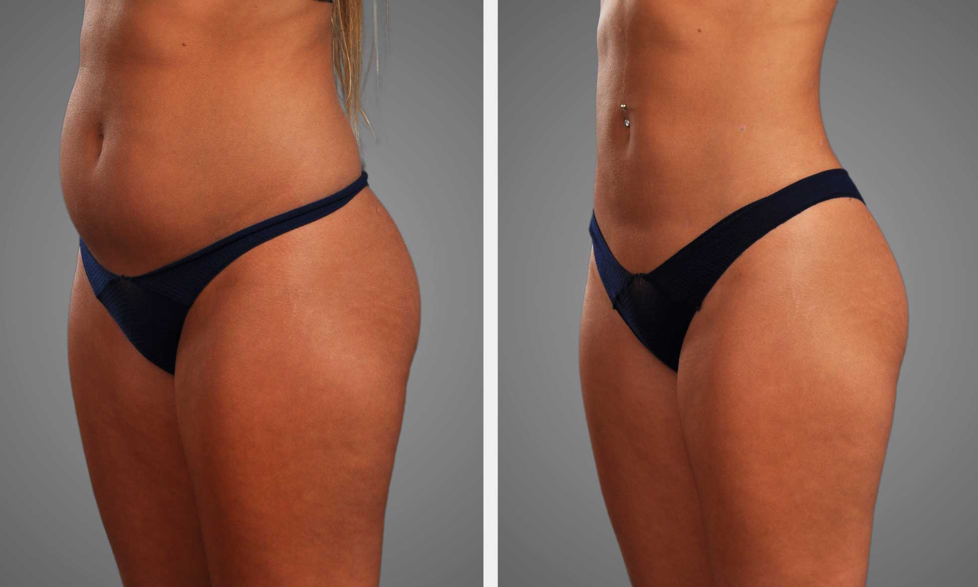
- Watch for asymmetry and contour irregularities of the buttocks post-BBL, as these could indicate fat necrosis.
- Fat necrosis is typically caused by poor blood supply, surgical trauma, infection, or underlying health issues. Therefore, pre- and post-operative care is important.
- Diagnosis usually includes physical examination, imaging tests, and occasionally a biopsy to verify the presence of necrotic tissue and direct treatment.
- Treatment can vary from conservative management and minimally invasive procedures to surgical correction, contingent upon severity and symptoms.
- Opting for a skilled and board-certified surgeon, adhering to aftercare instructions, and maintaining open communication with healthcare providers can minimize risks and enhance outcomes for patients undergoing BBL surgeries.
BBL fat necrosis symptoms often show as firm lumps, pain, or changes in skin color at the fat transfer site. Others may experience some swelling or a little knot that’s firm to the touch.
Fat necrosis treatment after BBL can consist of massage, warm compress, or minor surgery to remove the dead fat. Being aware of these symptoms and treatments assists individuals in identifying and addressing complications at an early stage.
Recognizing Fat Necrosis
Fat necrosis following a BBL is a potential complication in which the newly transferred fat cells perish as a result of inadequate blood flow. It may impact skin appearance, tissue consistency, and buttock contour. Early recognition of symptoms can spare patients unnecessary delays in care.
1. Skin Changes
Skin changes tend to be the early harbingers. You may observe bruising or discoloration that persists. The skin may appear red, purple, or even yellowish.
Other times, the skin is bumpy or uneven, dimpled or with hard patches. Swelling and warmth over the site might indicate inflammation or infection.
These symptoms may be mild or severe, but any persistent or worsening change should be evaluated by a physician. Any persistent changes, such as a patch of skin that remains dark or thick, could indicate fat necrosis.
2. Palpable Lumps
Lumps beneath the skin may be either soft or hard. Some feel like hard lumps; others are elastic. These lumps can be as small as a pea or much larger.
Most are painless, but a few cause tenderness upon palpation. They need to be aware that not all lumps are fat necrosis.
Others may be malignant or pre-malignant changes in the tissue. Fat necrosis lumps can even fluctuate in size or firmness.
The location plays a part as well, with deeper or larger lumps potentially indicating more serious necrosis. Doppler ultrasound can help show if there is blood flow in the mass or if it is dead tissue.
3. Pain and Tenderness
Pain is not universal. It can be a red flag. Others experience a mild soreness that intensifies with pressure or motion.
Sharp or lingering pain, particularly if it continues to increase, is not something you should overlook. Pain can be accompanied by swelling or even fever, which might indicate infection.
If it persists for weeks without getting better, this indicates continuing tissue damage and needs medical care.
4. Fluid Drainage
Leaking fluid from the incision or surgical site is a hallmark sign. The fluid may be yellow or cloudy, demonstrating dead tissue or infection. Occasionally, it is just a little damp, but other times, it is a gush.
The quantity and hue of the liquid are significant. Heavy drainage bogs down healing and might require special wound care.
It is essential to keep the area clean to prevent additional complications.
5. Asymmetry
Unevenness in the buttocks can form if fat necrosis is uneven. This can cause lumps, dents, or a misshaped overall appearance. Sometimes the changes are subtle and private.
Other times, they impact the fit of clothing or the way the body appears. Asymmetry can progressively worsen.
If you notice your form altering or one side appears uneven, it could signify the necrosis is spreading. This may impact satisfaction with surgery and require additional treatment.
The “Why” Behind It
Fat necrosis after BBL boils down to how fat cells react post-transplant. With BBL, surgeons harvest fat from one area, then inject it into the buttocks to provide more shape. It’s not an easy process. It requires talent, technique, and a taste for aesthetics, as not every fat cell makes it through the transfer. When fat cells don’t get enough blood flow in their new location, they perish. That’s the beginning of fat necrosis.
Without consistent blood flow, the transferred fat liquefies, leaving behind lumps, hard spots, or an uneven texture under the skin. Surgical trauma is a major factor in fat necrosis. Transporting fat is hard on cells. Even soft-spoken, considerate work can damage capillaries. Some regions might get bruised or crushed during the liposuction and fat injection steps.
The injury can be minor or severe, but regardless, it primes for suboptimal repair. If tissue is damaged during surgery, it is more prone to scar tissue or blood flow loss, increasing the risk of fat cell death. Post-operative infection and inflammation are a factor. When your body battles germs or inflammation, it pumps additional cells and fluids to the region.
This reaction can delay healing, impair circulation, or make the fat break down more quickly. Infection can manifest as redness, pain, or fever and can exacerbate fat necrosis. Even low-level swelling or a mild, difficult to detect infection can push it over the edge. These are the risks aftercare and maintaining surgical site cleanliness are crucial.
Patient factors count as well. Not everyone’s body responds in the same manner to a BBL. Problems such as diabetes, poor circulation, or a weakened immune system can delay healing. Smokers are at additional risk because nicotine constricts blood vessels, further restricting blood flow to new fat. Age, body weight, and adherence to aftercare all factor in as well.
If you have a history of poor wound healing or blood flow issues, you should be extra cautious and probably consider surgery twice. The surge in BBL procedures tracks beauty trends from the 90s “heroin chic” aesthetic to the social media and celebrity-driven curves of today. The choice to have a BBL is frequently influenced by peer pressure to conform or appear a certain way.
The operation is intricate and outcomes can be challenging to anticipate. Issues such as ‘diaper booty,’ lumps or hard spots can appear months or years down the line. Other research suggests that fat necrosis occurs in as much as 1 out of every 3,000 cases, a seemingly low number, but a worthy statistic for those considering surgery.
Balancing the allure of a new face with these dangers is crucial, and consulting candidly with an experienced surgeon aids in establishing realistic expectations.
Diagnosis and Confirmation
Detecting fat necrosis post BBL begins with a physical exam. Doctors examine the site for any subcutaneous lumps or bumps. These lumps might be initially soft or painful and then become hard and firm over a few weeks. Most manifest within the first three months post-surgery when the body is still adapting. If someone feels a bump that doesn’t subside after six or eight weeks, that’s a hint it might be fat necrosis rather than typical healing.
These lumps can vary in size and consistency. There are tiny ones that are barely felt and others that are larger and more prominent.
A physical exam is just the beginning. Of course, doctors diagnose with imaging. Ultrasound is popular as it is convenient and secure. It reveals what is happening beneath the surface and whether the tissue is viable. Occasionally, an MRI might be required for a nearer look, particularly if the lump is deep or difficult to detect by feel alone.
These scans capture specifics that differentiate fat necrosis from issues such as cysts or scar tissue. In certain locations, mammography is employed for fat necrosis that is adjacent to breast tissue, but for BBL, ultrasound and MRI are the standard.
If the lump persists or exhibits abnormal changes, a biopsy is next. This involves taking a small sample of the lump and examining it in the laboratory. A biopsy will determine if the tissue is necrotic fat, normal healing, or something else. This is key, as it can help eliminate other problems.
Take diagnosis and confirmation for instance; not every lump post BBL is fat necrosis. It may be scar tissue or, less commonly, infection. Receiving a definitive diagnosis allows for the appropriate therapies to begin immediately.
Symptom tracking is beneficial. They might have patients keep a pain diary, rating pain on a zero-to-ten scale and noting other changes. This assists physicians in correlating the symptoms to what they discover on exams and imaging.
Working in tandem, the patient and care team can identify trends that could be overlooked during an isolated appointment. Fat necrosis diagnosis is optimal when a multidisciplinary team of experts collaborate. Surgeons, radiologists, and at times pathologists all contribute their expertise.
This team approach makes the diagnosis more likely to be correct and the plan for care tailored to each case. Treatment begins with observation and conservative measures. If lumps persist or become painful, surgical excision or liposuction may be indicated.
Treatment Pathways
Treatment for fat necrosis post-BBL varies based on the severity and duration of symptoms. In most cases, we begin with conservative options. Sometimes more invasive procedures are necessary if there is persistent pain or distortion.
| Treatment Option | Description | Effectiveness |
|---|---|---|
| Observation | Monitoring without intervention | High for mild cases |
| Needle Aspiration | Removal of fluid or necrotic fat with a needle | Moderate; quick relief |
| Steroid Injections | Reduces inflammation in the affected area | Variable; may need repeats |
| Surgical Excision | Removal of dead tissue surgically | High for persistent cases |
| Reconstructive Surgery | Restores shape after large tissue loss | High, but more invasive |
Conservative Management
| Strategy | Indication | Timeline |
|---|---|---|
| Watchful Waiting | Mild, small lumps | Weeks to months |
| Pain Diary | Ongoing discomfort | Ongoing |
| Healthy Habits | All cases | Ongoing |
| Early Intervention | Lumps beyond 6-8 weeks | After 2 months |
Conservative management is most effective for mild symptoms. Most lumps subside or disappear in a matter of months. Typically, you begin with rest, gentle activity, and proper nutrition. This promotes natural healing and helps to prevent the likelihood of more severe issues.

Patients should maintain a pain diary using a zero to ten scale. They should note any symptoms such as swelling, warmth, or changes in skin color. This aids physicians in identifying problems early and determining whether additional intervention is necessary.
Pain is generally handled with OTC medication. Ice packs and mild massage are beneficial, but any acute changes mean a trip to the doctor. If lumps persist beyond 6 to 8 weeks post-surgery, they require additional workup.
Minimally Invasive Options
Needle aspiration can drain fluid that accumulates beneath the skin. It’s a less invasive approach and can provide fast relief from inflammation or stiffness. In some instances, we still utilize steroid injections to reduce the swelling and assist the area in healing.
Minimally invasive interventions suit moderate diseases. The key advantage is less recovery and risk than surgery. These alternatives can prevent scars or prolonged healing periods.
Each has its advantages and disadvantages. Aspiration and steroids might not repair substantial or persistent lumps. Complications such as infection or irregularities can occur, so patients should discuss options thoroughly.
Surgical Correction
Checklist for Surgical Correction:
- Full health check to spot any risks for surgery.
- Review surgical history, medicines, and allergies.
- Imaging or ultrasound to map out the necrosis.
- Definite route regarding how much and where to excise.
- Go over consent and talk about potential outcomes and the potential for additional surgery.
Surgery is used if other treatments don’t work or the necrosis creates significant issues. Other times, reconstructive surgery is required. Revision BBL is typically deferred until healing is established and stable, often three to six months post-op.
Aftercare involves wound inspections, infective prophylaxis, and activity resumption instructions. Recovery can take weeks to months, with ultimate results displayed up to six months.
Risk Factors
Fat necrosis post-BBL is not uncommon, and certain individuals are at increased risk. The risk of fat necrosis increases or decreases depending on health, habits, and surgical technique. Understanding what raises the risk assists patients and providers in making more informed decisions.
Age is a powerful one. Middle-aged women, around 50 on average, are at greatest risk. Those over 40 are in the high-risk category and typically require additional pre- and post-operative evaluations. This risk is increased in women with pendulous breasts, as changes in breast tissue can impede blood flow and fat survival.
Smoking is a big risk. Smoking impedes circulation and compromises the health of fat cells. Stopping before surgery helps a lot! Quitting smoking just a few weeks prior can result in approximately 75% better fat survival in the recipient site. This is an easy step that can significantly reduce risks.
Medical interventions such as brachytherapy or accelerated partial breast irradiation bear an obvious association with fat necrosis. Symptomatic fat necrosis develops in 1 to 50 percent of cases after these procedures. It depends on the amount of tissue exposed to the radiation and the intensity of the dose. Those who have had adriamycin-based chemo are also at risk, as this drug can damage healthy fat cells and impede healing.
Oncologic surgery patients—those who underwent surgery for cancer—are among the high-risk groups. These patients may have had altered tissue from previous treatments predisposing them to fat necrosis. They require closer monitoring and assistance post-BBL.
Underlying health issues make a big impact. Diabetes, for instance, can impede healing and reduce blood flow to fat grafts. Poor blood sugar control makes fat cells less likely to survive the transition. Other conditions that interfere with blood flow or the immune system may increase the risk as well.
How the surgery is performed is a significant concern. More experienced BBL surgeons generally have superior outcomes and less fat necrosis. Employing good technique, such as not applying too much fat at a time, selecting a proper location, and treating tissue gently, can reduce risk.
Patient care is key. Sticking to pre-op and post-op care rules can help. This includes not smoking, eating right, avoiding stress on the treated site, and maintaining all follow-up visits. By skipping instructions or checkups, patients are at a much higher risk.
The Surgeon’s Role
The surgeon’s role in fat necrosis prevention and treatment after BBL is critical to minimizing complications. Selecting an experienced surgeon with a good track record in fat transfer is essential. Expert surgeons understand fat handling, choose the optimal area to inject, and apply safe techniques. This reduces the risk of fat necrosis, which can cause pain, lumps, or hardening. Some of the best outcomes come from surgeons who work in clinics with excellent safety and patient care protocols.
It’s the surgeon’s responsibility to ensure patients understand what can go wrong with BBL. The surgeon should discuss risks such as fat necrosis, infection, or asymmetrical outcomes prior to surgery. This talk provides the patients a true understanding of what they should expect so there are no surprises down the road.
Surgeons should explain to patients how fat necrosis may present, such as hard lumps, swelling, or discoloration of the skin, and what actions to take if these symptoms arise. This candid dialogue enables patients to make informed decisions on their therapy and identify issues early.
A preoperative checkup is required to identify any health concerns that could increase the risk of fat necrosis. The surgeon looks for things such as previous health issues, smoking, or thin skin which can impact healing and fat survival. For smokers, surgeons typically recommend that they quit before and after surgery.
This reduces the chances of hypoxia in the tissues, a major culprit of fat necrosis post-transfer. For thin skin or in cases where a large size change is required, the surgeon may opt to perform the transfer in two stages or use less fat at a time. This can aid in preventing lumps or dead fat from developing.
Transparent, frank conversations between patient and surgeon are key. Patients should feel free to discuss any concerns or objectives, so surgeons have to address all questions and set realistic expectations for the outcomes.
If fat necrosis makes an appearance, the surgeon conducts a thorough examination to determine the extent of the issue. They may employ a lighted retractor to visualize and palpate the necrotic adipose tissue. Focusing on the dead fat could translate into incisions under your butt cheek, adjacent to a scar, or inside a tattoo, depending on where the issue lies.
Some can be addressed with massage or medicine, while others require surgery to excise the necrotic fat. For big or difficult cases, multiple surgeries or a rebuild is required.
Conclusion
Bbl fat necrosis symptoms treatment They require prompt medical examinations, not guesswork. Surgeons tend to catch these symptoms quickly and assist with stage implantation planning. Most cases require only basic management such as rest, massage, or short-term medications. Some require a little more, like a minor operation. Treating these symptoms as early as you notice them helps keep things simple and safe. If you have more risk, such as being a smoker or having health issues, maintain close contact with your care team. Open discussions and check-ins are a game changer. For any new bumps or pain post bbl, it is best to get it checked by a physician. Be vigilant, be inquisitive, and put your health ahead.
Frequently Asked Questions
What are common symptoms of fat necrosis after a BBL?
Typical symptoms are hard lumps, skin discolorations, tenderness or irregular surface at the site. Others might experience hard nodules or swelling. If you experience these symptoms, seek your surgeon.
How is fat necrosis diagnosed after a BBL?
Physicians usually diagnose fat necrosis by physical exam and history. Imaging tests, such as ultrasound or MRI, can assist in confirming the diagnosis.
Can fat necrosis heal on its own?
Mild cases of fat necrosis may resolve on their own over time. Persistent symptoms or discomfort should be evaluated by a medical professional for proper management.
What treatments are available for fat necrosis after a BBL?
Therapies can involve monitoring, massage, or antibiotics in mild instances. In select cases, surgical excision of the affected tissue may be required, particularly if the tissue is painful or infected.
What causes fat necrosis after a BBL?
Fat necrosis happens when fat cells that are transferred don’t get enough blood and shatter. This can be caused by overfilling, pressure on the area, or healing complications.
Are there risk factors for developing fat necrosis after a BBL?
Yes. Risk factors are large fat transfer volumes, poor blood circulation, smoking, and certain medical conditions. Adhering to your surgeon’s post-op care guidance minimizes the risk.
How can a surgeon help prevent or manage fat necrosis?
A good surgeon uses safe fat transfer techniques and clear aftercare. Early detection and intervention are key to minimizing complications and maximizing an outcome.