Key Takeaways
-
AI body scanning gives incredibly accurate, unbiased measurements that allow you to differentiate fat from muscle lost and personalize body sculpting plans.
-
Employ scan-driven 3D models and algorithmic analysis to personalize treatments, match modalities to needs, and adapt plans in real time.
-
Frequent scans allow for objective tracking of progress, clear before and after comparisons, and early detection of complications or plateaus.
-
Use scan data to analyze health indicators like visceral fat and muscle asymmetry, increasing safety and providing better long-term metabolic recommendations.
-
Combine scanning with consults to increase patient understanding, manage expectations, and empower practitioner decisions.
-
Take follow-up actions such as periodic scans, recording baseline and final measurements, and adjusting nutrition or exercise plans based on algorithmic recommendations.
AI body scanning before body sculpting procedure is a digital assessment that maps body shape and tissue layers to guide treatment plans.
It provides precise measurements, tracks changes over time, and helps clinicians choose device settings and target areas. The scans reduce guesswork and can improve consistency in results across sessions.
Patients receive visual reports and progress data that support informed decisions and realistic expectations before any procedure.
AI-Powered Enhancements
AI body scanning with BodyBlock introduces an additional level of accuracy and insight prior to body sculpting procedures. It produces a 360-degree true three-dimensional model of the body and generates information that helps doctors design, adjust, and monitor therapies with less speculation and less estimation.
1. Unmatched Precision
AI-powered 3D scanners like Styku and ShapeScale provide millimeter-level precision, usually within approximately 9 mm, for external shapes and surface markers. This granularity aids in identifying minor changes in fat layer thickness and muscle mass that standard tape measures or scales overlook.
Scans generate objective maps of hard-to-lose fat deposits, enabling a provider to designate precise target areas for targeted fat-melting energy or lipolysis. Repeatable, consistent scans minimize human error in measurement and make longitudinal tracking reliable. Practitioners can compare scans week to week and see objective change rather than rely on subjective photos.
2. Tailored Planning
AI-based enhancements utilize the 3D model and composition data to create personalized treatment plans that identify specific body types and objectives. They can include modality decisions, such as cryolipolysis for more superficial pockets, radiofrequency for skin tightening, or focused ultrasound for deeper fat, depending on measured tissue depth and skin laxity.
Real-time feedback during a session allows clinicians to adjust energy settings or handpiece placement as the scan reveals tissue response. Your waist, the contour of your thighs, or loose skin in particular become quantifiable goals in the plan, not nebulous objectives.
3. Predictive Outcomes
Algorithms mine previous pre- and post-procedure data and predict expected outcomes for a new patient with comparable statistics. We then used those models to establish achievable expectations for fat reduction percentages, possible muscle definition improvements, and probable contour alterations.
They highlight trends that could signal increased complication risk, like high visceral fat or abnormal physiques. Advanced visualization tools paint predicted changes onto the patient’s 3D model, assisting in goal alignment and informed consent.
4. Objective Tracking
Routine scans measure fat proportion, muscle mass, and waist-to-hip ratios and package that into easy-to-understand reports and charts. Clinicians can present side-by-side before-and-after scans to confirm treatment efficacy and to bolster nutrition and exercise compliance.
These data capture changes across numerous composition factors, usually 14 prime markers, so improvements are wide and detailed, not a single measure.
5. Enhanced Safety
Pre-procedure AI screening detects health risks such as high visceral fat or abnormal BMI and helps determine candidacy for invasive approaches. Detailed scan data inform surgical planning and minimize intraoperative surprises.
Post-procedure follow-up scans can spot early signs of uneven healing or complications and inform timely intervention.
The Technology
These AI body scanning systems marry precise hardware with machine learning to map the body to millimeter detail, generating a baseline for body sculpting. Such systems input data into treatment flows, support medical weight loss programs, and assist doctors in selecting energy types, cooling levels, and treatment targets with confidence.
Data Capture
|
Category |
Example Measurements |
|---|---|
|
Linear |
Height, limb lengths, waist circumference (cm) |
|
Volumetric |
Limb volume, trunk volume (liters) |
|
Surface |
Skin fold thickness, surface area (cm²) |
|
Spatial |
Posture angles, joint alignment (degrees) |
|
Tissue |
Estimated fat distribution, muscle contours |
Scanners such as Styku and ShapeScale employ infrared, structured light or depth-sensing cameras to capture these points quickly. Full body 3D scans provide a holistic overview of shape and posture rather than focusing on specific regions.
Archiving past scans builds a timeline of long term change. Clinicians are able to export tables and lists of measurements for progress reports or link data to weight management programs to update caloric targets and exercise plans.
Practitioners rely on scan outputs to construct detailed treatment maps. For instance, a clinician can highlight spots with more subcutaneous fat and plan combination treatments. Radiofrequency is used for deeper warming and cryolipolysis is used for surface-level fat according to the scan profile.
Algorithmic Analysis
AI scans the raw scan data and flags trends like consistent fat loss in the abdomen or unexpected flank asymmetry. Algorithms measure muscle gain compared to fat loss, providing percent-change metrics that are simpler to take action on.
Automated alignment between scans accelerates evaluation of therapy response. It can draw attention to more subtle enhancements, like a 5% decrease in subcutaneous thickness or a pose shift that changes contours.
Predictive models suggest personalized adjustments. Tweak RF intensity to reach 15 to 20 mm penetration for immediate tightening. Reduce cooling when tissue temperature approaches lower safety bounds. Add regenerative steps like adipose-derived stem cell therapy to aid healing and skin quality.
AI assists in identifying muscle tone imbalances which require focused physiotherapy or resistance training to optimize symmetry in advance of sculpting procedures.
3D Visualization
Interactive 3D models allow patients to rotate and zoom into particular areas, view underlying fat and muscle maps, and compare projected results side by side. Before-and-after renderings utilize the same scan grid for a precise visual comparison.
Visual feedback fuels motivation and compliance. By showing probable outcomes and bounds, it diminishes unwarranted optimism and aids in confirming treatment directions.
Clinicians can superimpose treatment zones, cooling, and energy type on the model for transparent planning. Real-time monitoring during sessions, guided by the scan model and data from the FDA-cleared device, fine-tunes delivery and enhances safety, minimizing side effects and trimming down the total treatment time.
Beyond Aesthetics
AI body scanning prior to body sculpting offers more than a picture — it offers clinical information that connects appearance to wellness. Scans establish a baseline for focused treatment, allow doctors to monitor progress, and facilitate coordinated plans that incorporate diet, fitness and monitoring.
Beyond aesthetics, here are some fundamental health indicators and how they inform safer, more sustainable care.
-
Body composition breakdown: precise measures of fat mass, lean mass, and bone mass help prioritize treatments and set realistic goals.
-
Visceral fat estimation: Volumetric or attenuation metrics show harmful abdominal fat levels tied to cardiometabolic risk.
-
Regional fat distribution maps subcutaneous versus visceral fat across the trunk and limbs. This information is used to direct treatments and forecast contouring response.
-
Muscle mass and quality localized lean tissue measures reveal atrophy or hypertrophy and help plan toning or strength work.
-
Muscle asymmetry indices: Side-to-side comparisons quantify imbalances that affect posture, gait and injury risk.
-
Posture and alignment markers: Three-dimensional positional data can flag spinal or pelvic tilt that changes surgical planning or exercise choice.
-
Metabolic proxies, such as lean mass and fat ratios, inform basal metabolic rate estimates and energy needs.
-
Skin and soft-tissue thickness: Thickness maps affect device selection and predict response to energy-based therapies.
-
Progress tracking metrics: Repeat scans provide objective longitudinal data for outcomes, safety, and patient counseling.
-
Risk stratification factors: Combined markers help clinicians assess suitability for procedures and integrate medical weight loss when needed.
Visceral Fat
Visceral fat increases risk of type 2 diabetes, hypertension, and dyslipidemia. It raises inflammation markers linked to cardiovascular disease. Additionally, it correlates with insulin resistance and fatty liver disease.
Visceral fat is also associated with higher overall mortality in long-term studies. Aim to reduce visceral fat and surface-level fat with your interventions. Scan to track visceral fat in medical weight loss or body sculpting programs so clinicians can modify treatment.
Scan data assists in patient education on health benefits from reducing visceral fat, fueling sustainable lifestyle transformation and enhanced self-image exhibited in long-term results.
Muscle Asymmetry
Go beyond aesthetics and uncover muscle mass imbalances between limbs and areas with detailed scan results. Custom-fit exercise and toning plans address asymmetry, including unilateral strength work and targeted physical therapy.
Measure changes over time to document actual progress in symmetry, which helps increase patient happiness. Early correction reduces injury risk and enhances functional performance. Scans can inform the integration of exercise into holistic treatment plans.
Metabolic Insights
Go beyond aesthetics. Body composition estimates metabolism and daily calorie requirements. Shifts in lean mass alter energy consumption.
Tweak nutrition and exercise based on scans revealing lean mass loss while cutting to protect metabolism. Give metabolic profiles at medical weight loss consults for long-term wellness.
AI-generated insights can alert when to incorporate combination treatments, which enhance efficacy and safety profiles over single approaches.
The Patient Experience
AI body scanning prior to body sculpting provides patients a definitive, trackable baseline from which to monitor transformation. A quick description of where scans occur in the care pathway goes a long way to establishing expectations before diving into the visit stages underneath.
Initial Consultation
Baseline 3D scans capture body shape, circumferences and composition without touch. The scans require minutes, are non-invasive and generate images and metrics that display fat distribution, muscle mass and symmetry. Clinicians leverage that data to establish achievable targets and to delineate treatment zones with precision, such as addressing subcutaneous fat deposits at 2 to 4 cm depth or a weak gluteal quadrant for muscle-tone devices.
AI recommends probable working modalities by matching scan patterns to device profiles, such as cryolipolysis for focal fat bulges, radio frequency for skin tightening, or electromagnetic stimulation for muscle. Outcomes and choices are presented graphically so patients can contrast possible results and choose a strategy that matches lifestyle and finances.
Documenting metrics, including waist, hips, limb volumes, and body-fat percentage, delineates a definite baseline for comparison down the road and for shared decision making. Personal comfort is scheduled too. Therapies like immersive VR or custom playlists can minimize anxiety during scans and post-treatment.
Clinics can provide dimmable lighting, temperature control, and aromatherapy to make that inaugural visit serene and personal.
Progress Monitoring
Routine follow-up scans give you objective marks of transformation and eliminate the guessing process. Scans every four to eight weeks fit with many treatment cycles and allow clinicians to witness fat reduction, volume shifts, or gains in muscle tone in a metric form. If scans plateau, clinicians can adjust device settings, swap frequencies, or incorporate complementary therapies.
Real-time monitoring tools and pain-feedback systems can guide immediate modifications to sessions, enhancing comfort and safety. It’s reinforcing to show patients side-by-side photos and number trends to remind them of their progress and keep their motivation high.
Short milestone reports, badges, or simple charts can help patients adhere to exercise, dietary, or maintenance plans. Immersive breaks or guided meditation provided during treatment visits prevent anxiety and make return visits smoother.
Final Assessment
|
Metric |
Baseline |
Final |
Change |
|---|---|---|---|
|
Waist circumference (cm) |
92 |
86 |
−6 |
|
Body fat (%) |
31.2 |
|
|
|
27.8 |
−3.4 |
|
|
|
Thigh volume (cm3) |
8,200 |
7,750 |
minus 450 |
Final scans contrast results right back to original objectives and illustrate transformations in contour, fat ratio, and muscle tone. A brief summary report identifies targets hit, targets missed, and maintenance suggestions.
Education materials describe risks, benefits, and next steps so patients leave knowledgeable and prepared to navigate long-term outcomes.
Practitioner Perspective
AI body scanning serves as an objective bridge between consultation and treatment. It provides a definable, quantifiable baseline and a common point of reference for clinician and patient alike. The scans flow into treatment planning, documentation and follow-up, and they simplify incorporating nutrition, exercise and adjunct therapies in a unified care pathway.
Consultation Tool
3D scans reveal surface contours, volume fluctuations, and asymmetries like nothing photos can. Through the practitioner lens, side-by-side renderings help practitioners set realistic goals and avoid misconceptions about what is likely to occur.
Displaying a simulated post-treatment image allows patients to consider their choices and consent with greater clarity. Recommendations are driven by scan data. Fat distribution maps, circumference measures, and regional volume estimates support data-driven decisions between fat reduction, RF tightening, or hybrid approaches such as PRP-assisted contouring.
Paired with genetic markers or biomarker reports, the scan data can inform more personalized plans. Pictures make it easier to explain to other teams. Nurses, therapists, and trainers all see the same images and metrics to coordinate prehab or post-protocol care.
This common record fosters faith. Patients watch the clinic deploy precise instruments, not empty slogans. With scans and AI models guiding the consult, you’re communicating an intention towards precision. That credibility counts for nervous patients and for those who are provider shopping in a competitive marketplace.
Treatment Validation
Scans provide pre- and post-measures that outperform pictures. From a practitioner perspective, they measure fat mass decrease, muscle tone indicator changes, and skin surface texture changes over time.
Hard numbers enable clinics to verify what machines and protocols work optimally. For instance, do next-gen fat freezing or intelligent RF systems deliver better results? It’s about the data that’s tracked versus the feeling. That data backs protocol polishing.
Keeping scan records supports compliance and quality assurance. Accurate logs of treatment parameters and measured outcomes satisfy audit requirements and support clinical decision-making. When results lag, scan histories illuminate where protocol modifications or adjuncts such as PRP or medical weight-loss referral are indicated.
Scans guide long-term strategy. If data demonstrate improved retention when paired with exercise physiology programs, practitioners can establish formalized co-pathways.
Client Education
Create a simple checklist: hydrate, avoid smoking, follow targeted exercise, keep protein intake steady, and attend follow-ups. Show the dos and don’ts visually, connected to the scan areas the client is looking to modify.
Explain body composition in plain terms: fat versus lean mass and how each affects shape and health. Use scan images to illustrate where diet and strength work will assist and where device-based contouring is necessary.
Give clear tips: short resistance sessions, protein timing, and gradual caloric shifts, along with relaxation or distraction programs to lower treatment anxiety during sessions.
Provide progress reports. Small, quantified successes from scan data empower compliance and patient belief, which numerous practitioners say result in improved health.
Future Outlook
AI body scanning will propel body sculpting into an era in which the planning, delivery and follow-up are a single joined process. Scanners that now generate three-dimensional maps will connect to databases containing results from thousands of patients. This will allow systems to compare a new patient’s shape and metabolism to similar cases and recommend targeted treatment courses. Imaging will encompass not only surface shape but also tissue composition and metabolic markers, providing a more comprehensive understanding of fat distribution and response. That richer information will allow doctors to choose the optimal mix of tools, energy levels, and session timing for each patient.
Look for hardware and chemical breakthroughs to partner with AI. Researchers are trialing targeted nanoparticle delivery and fat cell–acting drugs. Combined with precise scans, those agents can be directed at specific areas to increase effectiveness and decrease side effects. Imaging and monitoring will increase in detail, with real-time tissue temperature maps and continuous vital-sign feeds connected to the scanner and control software. Systems can automatically halt energy delivery if temperature or perfusion deviate from safe ranges. This may make treatment sessions safer and more consistent.
Anticipate far greater accuracy and customization. AI models will harness body composition analysis, previous results and demographics to calibrate energy levels and select modalities aligning with skin type, fat distribution and metabolic profile. For instance, a patient with denser subcutaneous fat may receive an alternate radiofrequency protocol than one who has looser connective tissue. Treatment plans will be customized to probable tissue reaction, not merely to broad body area. That cuts down on trial and error visits and compresses care timelines.
Wider use across wellness and cosmetic dermatology will occur as tools become easier and interoperable. Clinics will connect scanners, treatment platforms, and electronic records so a dermatology practice can do scans, pull AI suggestions, and provide non-invasive contouring with built-in safety protocols. This distribution will be aided by patient care innovations like built-in cooling, vibration, topical anesthetic distribution, and live pain tracking that modifies parameters during therapy. Those comforts help make processes more permissible and available to more patients.
Better results and happier customers will come from constant innovation and data feedback loops. With continual outcome tracking, AI will improve predictive algorithms and distinguish which protocols provide lasting change for which phenotypes. Treatments will become even faster, with fewer sessions and more consistent long-term outcomes. Safety will advance with protocols incorporating ongoing vital monitoring, auto shutoff for safety signs, and fine-tuned temperature control.
Conclusion
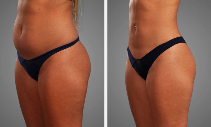
Here’s where AI body scans bring obvious value to body sculpting. They scan fat and muscle with precision. They assist in strategizing safe incisions, targeting symmetry outcomes and monitoring recovery. Patients get clearer expectations and proof of transformation. Surgeons get exact guides and reproducible data. Clinics reduce rework and accelerate follow up. Real cases surprise less and the results hold more steady.
Use simple checks: compare scans before and after, note exact volume shifts, and log photos with measurements. Little things like that make care more dependable. If you’re considering it, request scan samples and a data review. Contact your clinic to view sample scans and discuss how they suit your plan.
Frequently Asked Questions
What is AI body scanning before a body sculpting procedure?
AI body scanning utilizes cameras, sensors, and software to generate an accurate 3D model of your body. It maps your curves, fat placement, and symmetry to inform planning and results.
How does AI improve treatment planning?
AI then analyzes the 3D model to make recommendations about target areas, volumes, and treatment patterns. This enhances accuracy, minimizes random estimations, and aids in establishing expectations.
Is AI body scanning safe and private?
Yep, scanning is non-invasive and fast. Clinics ensure data protection law and secure storage. Inquire regarding encryption and duration of image storage.
Will AI scanning predict my exact results?
No. AI gives very precise simulations and probabilities, but outcomes still shift with healing, lifestyle, and technique. Simulations are guiding aids, not promises.
How does AI affect recovery and complication risk?
AI can reduce overtreatment and enhance symmetry, thereby reducing complications. It does not substitute for surgical expertise or post-op care.
Do all practitioners use AI body scanning?
No. Adoption differs by clinic, region, and procedure. Question providers on their technology, experience, and how they incorporate scans into planning.
How should I choose a clinic that uses AI scanning?
Seek board-certified clinicians, tracked results and open data policies. Ask to view some before and after cases and how the AI informed the plan.