Key Takeaways
- Lipedema is a chronic, progressive fat disorder that affects the legs and sometimes arms and is commonly misdiagnosed as obesity or lymphedema. Clinicians should look for symmetry, pain, and sparing of the hands and feet when suspecting lipedema.
- Best diagnostic methods for lipedema A systematic diagnostic pathway enhances accuracy by integrating a thorough history and physical examination with imaging such as MRI and ultrasound, along with selective blood work to rule out metabolic or endocrine etiologies.
- Differential diagnosis is critical to prevent misdiagnosis. The best diagnostic methods for lipedema use clear exclusion criteria and comparison charts to differentiate lipedema versus obesity, lymphedema, and venous disease.
- Early detection and standardized checklists lead to better outcomes. Screen the most at-risk patients, record symptoms through diaries or digital tools, and inform primary care about early manifestations.
- New diagnostic tools such as genetic markers, AI-powered imaging analysis and sophisticated non-invasive scans hold potential for more accurate and less invasive diagnosis and should be adopted into research and clinical care when accessible.
- Once diagnosed, pursue an individualized management plan that incorporates conservative therapies, multidisciplinary specialist referrals, ongoing monitoring and psychosocial support to maintain function and quality of life.
What are the best diagnostic methods for lipedema: clinical exam, patient history, and imaging tests like ultrasound and MRI.
Diagnosis is based on lipedema fat distribution, pain, and swelling in conjunction with eliminating lymphedema via objective testing.
Coupling appearance scales to limb volume provides more accurate diagnostics.
Early, definitive diagnosis directs treatment decisions such as compression, therapy, or surgery and minimizes misdiagnoses from other specialists.
The main body breaks down every method, step, and proof.
Understanding Lipedema
Lipedema is a long-term disease characterized by irregular, tender fat deposits, predominantly in the legs and occasionally arms. It’s a different disease than standard obesity and different than lymphedema, though symptoms intersect. Recognizing the triad of symmetrical swelling, tissue that bruises easily, and disproportionate limb fat distribution is key to the diagnosis.
Risk factors are heritable or genetic, hormonal changes, and being female. It typically begins during puberty, pregnancy, menopause, or other significant hormonal changes.
The Condition
Understanding Lipedema is a progressive disease with disproportionate fat distribution that can exacerbate over time. The tissue changes are not lifestyle caused. Diet and exercise seldom undo the abnormal fat deposits.
With lipedema, individuals usually continue to gain or lose weight in other areas, but the affected limbs remain stubbornly impervious. Symptoms generally begin or worsen during hormonal milestones. Puberty is common, but pregnancy and menopause are also significant.
Over time, the additional tissue can restrict joint range and cause actual mobility issues. Other research suggests defective lymphatic transport of fat tissue results in local hypoxia and inflammation, which potentially fuels progression. Family history helps; roughly 15% report a relative with similar signs.
About 11% of women have lipedema, but underdiagnosis probably makes the actual figure higher.
The Symptoms
Classic symptoms are symmetrical swelling of the legs, tenderness, easy bruising, and pain in the affected limbs. Unlike generalized edema, the hands and feet are spared, which helps distinguish lipedema from lymphedema.
They frequently complain of heaviness, chronic fatigue, and difficulty walking extended distances. The skin and subcutaneous layer can feel soft, nodular, or rubbery. In addition, fluid that pools from gravity can still cause ankle or foot swelling in some people, even when blood vessels and lymphatics are normal, complicating the clinical picture.
Easy bruising with minimal trauma and disproportionate leg fat compared with the trunk are classic symptoms that clinicians search for.
The Stages
Stage 1 presents as smooth skin with enlarged subcutaneous fat and edema. All of these changes can be subtle and easy to overlook.
Stage 2 is characterized by an uneven skin surface, lumpy nodules in the fat, and further increase in size. Stage 3 has larger fat deposits, lobular deformities, and more limited mobility.
Stage 4 may involve secondary lymphedema, extreme joint pain, and significant disability.
| Stage | Clinical features |
|---|---|
| 1 | Smooth skin, fine nodularity, mild enlargement |
| 2 | Uneven surface, palpable nodules, bruising |
| 3 | Large deforming fat pads, mobility limits |
| 4 | Secondary lymphedema, severe pain, disability |
Compression garments can help alleviate pain, reduce inflammation and prevent fluid retention. Stage dictates benefit.
The Diagnostic Pathway
The diagnostic pathway for lipedema transitions from clinical suspicion to confirmation with history, exam, focused testing and ruling out other conditions. Early steps set the stage: clinicians gather detailed history and perform a focused physical exam, then add imaging and specialized tests as needed.
Since diagnosis is frequently delayed, sometimes for a decade, and missed on initial contact, a pathway cuts down on misdiagnosis and the risk of improper counseling such as exclusively lifestyle change.
1. Clinical Evaluation
Take a detailed history, including timing and progression of the symptoms, as well as any familial occurrence of limb fat. Just note age at first change. Many patients receive their diagnosis after age 40, and delay is rife.
Inquire about pain, easy bruising, and responsiveness of limb size to diet and exercise. Record previous diagnoses and treatments, including weight-loss efforts that didn’t budge disproportional limb fat.
Conduct a diligent physical examination. Check for symmetrical, bilateral fat deposition on hips, thighs, and sometimes arms, with relative sparing of hands and feet. Palpate tissue for softness, nodularity, and tenderness.
Measure limb circumference and compare sides. Observe skin and note pitting edema. Quantify functional thresholds, like not being able to get up from the floor or get clothes on.
Document the patient’s pain and how it affects their life. Employ standardized instruments such as visual analog scales, limb volume charts, and quality-of-life questionnaires to maintain consistency between visits and clinicians.
2. Differential Diagnosis
Distinguish lipedema from obesity, lymphedema, and chronic venous disease. Obesity leans towards generalized weight gain and responds more robustly to weight loss. Lipedema demonstrates disproportionate extremities and a lack of response to dieting.
Lymphedema may present with unilateral swelling, pitting edema, and foot involvement, which are unusual in lipedema. Venous insufficiency can lead to skin changes and varicosities instead of the nodular fat seen in lipedema.
Use exclusion criteria to exclude from endocrine or metabolic. Use a simple comparison chart listing features: symmetry, foot sparing, pain, compressibility, and response to weight loss.
This graphic helps clinicians and patients understand why diagnosis varies.
3. Imaging Techniques
Employing MRI to measure fat distribution and tissue sodium helps differentiate lipedema from other conditions, giving high-resolution fat layer maps. Ultrasound provides a low-cost alternative to demonstrate increased subcutaneous fat thickness and typical fibrotic alterations.
Magnetic resonance lymphangiography visualizes lymphatic vessels and may demonstrate vascular overload or subtle edema not evident on conventional imaging. Always read the imaging in the context of your clinical exam to avoid relying too heavily on one modality.
4. Specialized Tests
As mentioned earlier, when history suggests endocrine etiologies, order lab tests to rule out thyroid, adrenal, and metabolic disorders. Consider tissue biopsy if imaging and exam are inconclusive.
Histology can demonstrate adipocyte hypertrophy and fibrosis. If possible, participate in or refer to biorepositories to bolster research efforts and gain access to cutting-edge testing.
Keep a compiled list of accessible specialty tests and local referral centers to simplify care for patients frequently brushed off or stuck in diagnostic limbo.
Diagnostic Challenges
Lipedema is often unrecognized for years because its symptoms overlap with common conditions and clinical pathways aren’t clear. The classic presentation — bilateral, symmetrical, disproportionate fat on extremities sparing hands and feet, tissue tenderness, tightness, easy bruising and worsening during the day — should set off alarm bells. However, many providers simply don’t see it enough to notice the constellation.
Imaging may assist, but scans are primarily employed to exclude other concerns instead of being regular diagnostic tools.
Misdiagnosis
Lipedema is often misdiagnosed as either simple obesity or lymphedema. Obesity-centered guidance such as calorie restriction or weight-loss programs often doesn’t adjust the disproportionate limb fat. When lymphedema is later diagnosed instead, patients may be provided compression or decongestive therapies that overlook the root issue.
Misdiagnosis postpones efficient control and can allow symptoms to intensify. Unchecked progression causes increased tissue fibrosis and, consequently, more pain and functional restrictions. For most, years elapse between initial symptoms and accurate diagnosis, hampering efforts at early intervention like conservative care, focused physiotherapy, or surgery when appropriate.
To be misunderstood by providers inflicts obvious psychological damage. Patients say they feel dismissed, blamed, or stigmatized, which erodes trust in care and decreases help-seeking. Your solution involves clinicians utilizing specific diagnostic criteria, charting hallmark symptoms, and treating patient accounts of pain and bruising with respect to reduce mis-labeling.
Guideline Gaps
Unfortunately, there are no standardized diagnostic criteria for lipedema. Regional and provider-level variability is wide. Some specialists follow structured staging and typology systems while generalists apply intuition or treat it as obesity-related.
This lack of consensus affects patient care directly. Without evidence-based protocols, referrals are inconsistent, access to appropriate therapy is uneven, and research outcomes remain hard to compare. Listing gaps shows where work is needed: standardized staging, agreed measurement methods for tissue volume, clear imaging indications, and consensus on when to refer for surgical assessment.
Creating these protocols would help reduce practice variation and assist training programs. It would further render data across centers comparable and aid in a more precise definition of prevalence and course of disease.
Early Detection
Create a practical checklist to use in primary care and clinics:
- Family history of limb fat disproportion
- Bilateral symmetric limb fat with cuff phenomena (hands and feet spared)
- Daily worsening of heaviness
- Tenderness to touch
- Frequent easy bruising
Add red flags:
- Rapid functional decline
- Progressive fibrosis
Screen high risk groups regularly, especially with family history or early onset limb changes. Educate primary care teams to identify core signs and record them systematically, so referrals and diagnostic imaging are focused and timely.
Employ straightforward imaging such as ultrasound selectively to quantify subcutaneous tissue and exclude other etiologies. CT or MRI is reserved for complicated cases.
The Human Element
They are patient experience informs every stage of precise lipedema diagnosis. We need to understand how symptoms manifest, how patients describe them, and how doctors react. The human side is trust, validation, and ongoing support. These can be the difference between a patient falling from misdiagnosis into the abyss or emerging into the sunshine of appropriate care.
Patient Experience
- Challenge of getting doctors to acknowledge lipedema as a legitimate pathological disorder.
- Repeated advice to “lose weight” despite disproportionate fat distribution.
- Pain, easy bruising, and sensitivity that affects daily life.
- Barriers to specialist referral and diagnostic imaging access.
They manifest in visible symptoms that impact self-esteem and social life. They tell us they’re skipping parties, hiding their limbs in clothing, and restricting the activities that reveal their bodies. The tension reflects in career decisions and romantic affairs.
Too many patients become advocates. Patient groups share stories: one woman described crying with relief on receiving a diagnosis after years of dismissal. Some create online communities to advocate for improved care, gather resources and lobby for insurance coverage.
These stories can direct clinicians about practical effects and priorities for treatment. Go back to the first bullet list when designing the clinic and training staff to keep the patient concerns front and center. Leverage it to inform clinic hours, referral paths, and patient education materials.
Symptom Reporting
Maintain detailed logs of symptoms and triggers and track how things vary over time. Notice pain level, swelling patterns, and what aggravates or relaxes. Note down dates and events associated with flare ups.
Be candid about pain, range of motion restrictions and what activities have become difficult. Explain how symptoms disrupt your work, parenting or working out. Accurate terminology assists clinicians in differentiating lipedema from obesity or lymphedema.
Describe patterns: bilateral, symmetric limb enlargement, sparing of hands and feet, and sensitivity that differs from simple fat gain. Account for botched weight-loss attempts, particularly in your teens, and any track record of disordered eating. As high as 18% may establish chronic eating disorders after years of weight-failure cycles.
Use symptom diaries or apps for accuracy. It’s much easier to take advantage of digital tracking, which lets trends be quickly shared with providers and enables remote monitoring.
Psychological Impact
- Think of counseling, peer support groups, cognitive behavioral therapy, mindfulness apps, and crisis hotlines.
- Suggest referral to mental health professionals experienced with chronic illness.
- Add peer-led education and advocacy groups for social support.
- Promote physio and pain management programs featuring psych care.
Lipedema increases anxiety, depression, and social isolation risks. The prevalence of depression ranges from 31 to 59 percent. Uncontrolled pain, in particular, tends to feed brainworm.
Quality of life is frequently middling even with significant blows. The absence of diagnosis and treatment compounds the emotional burden. Mental health care must be included in any diagnostic trajectory to minimize long-term damage.
Future Diagnostics
Future diagnostics of lipedema are about earlier, more accurate detection and patient-friendly approaches. Research integrates genetic analysis, neuroimaging, AI, and mobile technology to distinguish lipedema from overlapping conditions, enhance diagnosis and monitoring, and inform precision treatment.
Genetic Markers
Future Diagnostics, continuing research, genetic identifiers for lipedema. Studies have indicated PIT-1 mutation and NSD1 gene alterations such as microdeletions. Identifying dependable markers would allow doctors to test family members who are at risk and detect the disease before clinical symptoms manifest.
If we can identify a panel of sensitive and specific genetic markers, it may allow targeted therapies based on a patient’s molecular profile. That might imply earlier lifestyle or clinical interventions and improved trial design for novel pharmaceuticals.
Family screening by genetic risk might be offered where mutations are well validated, though ethical and counseling steps are required. The table below lists markers now being investigated and their suggested connections.
| Genetic Marker | Type of Change | Proposed Link to Lipedema |
|---|---|---|
| PIT-1 | Mutation | Hormone regulation; possible role in fat distribution |
| NSD1 | Mutations / Microdeletions | Chromatin regulation; may affect adipose tissue growth |
| Candidate loci (various) | SNPs | Under investigation for risk modulation |
Artificial Intelligence
AI can scan imaging and clinical data to identify subtle patterns that clinicians overlook. Machine learning models trained on labeled MRI and ultrasound images are promising for distinguishing lipedema from lymphedema and simple obesity.
Others utilize deep learning to assign weights to features such as fat layer thickness, fibrotic changes, and vessel patterns. They can at least flag borderline cases and minimize inter-observer variability. Initial research indicates AI might reduce time to diagnosis and reduce false positives.
Their initial AI use cases include automated segmentation of fat compartments on MRI, pattern recognition in ultrasound speckle texture and decision-support tools that combine symptom scores with imaging. Broader datasets and external validation are required prior to typical use.
Non-Invasive Tools
Advanced MRI and high-resolution ultrasound are important non-invasive modalities. MRI can map fat distribution and demonstrate typical sparing patterns. Ultrasound can evaluate tissue compressibility and nodularity. Each is safer and more comfortable than biopsies.
Non-invasive tools minimize risk and enable frequent monitoring. Handheld ultrasound could deliver point-of-care evaluation in the clinic or the community, improving availability in locations without expert imaging.
Comparing options assists clinics in selecting tools by expense, accessibility, and diagnostic return. Future diagnostics involve new biomarkers and imaging protocols that seek to increase sensitivity and specificity so clinicians can depend less on exclusion and more on direct indicators of lipedema.
Post-Diagnosis
Once you get a confirmed lipedema diagnosis, immediate and defined next steps prime the path toward improved symptom management and quality of life. Our first steps are a full baseline work-up, documenting your staging and type, discussing your goals, and educating you on treatment options.
Keep an activity index, such as step counts from a phone app, and baseline pain, mobility, and mental state to inform later decisions.
Management
Conservative treatments are the cornerstone. Central to CDT is manual lymphatic drainage, compression therapy, structured exercise therapy, and skin care. Compression garments must be fitted and reviewed regularly.
Manual lymphatic drainage can decrease fluid swelling and is taught by specialists. Physical therapy works on strength, range of motion, and low-impact aerobic exercise to maintain mobility. Diet changes may alleviate symptoms.
Refer to a psychologist when disordered eating is present so dietary work is safe. Surgical options are for patients that do not get sufficient relief from conservative care or who have progressed disease.
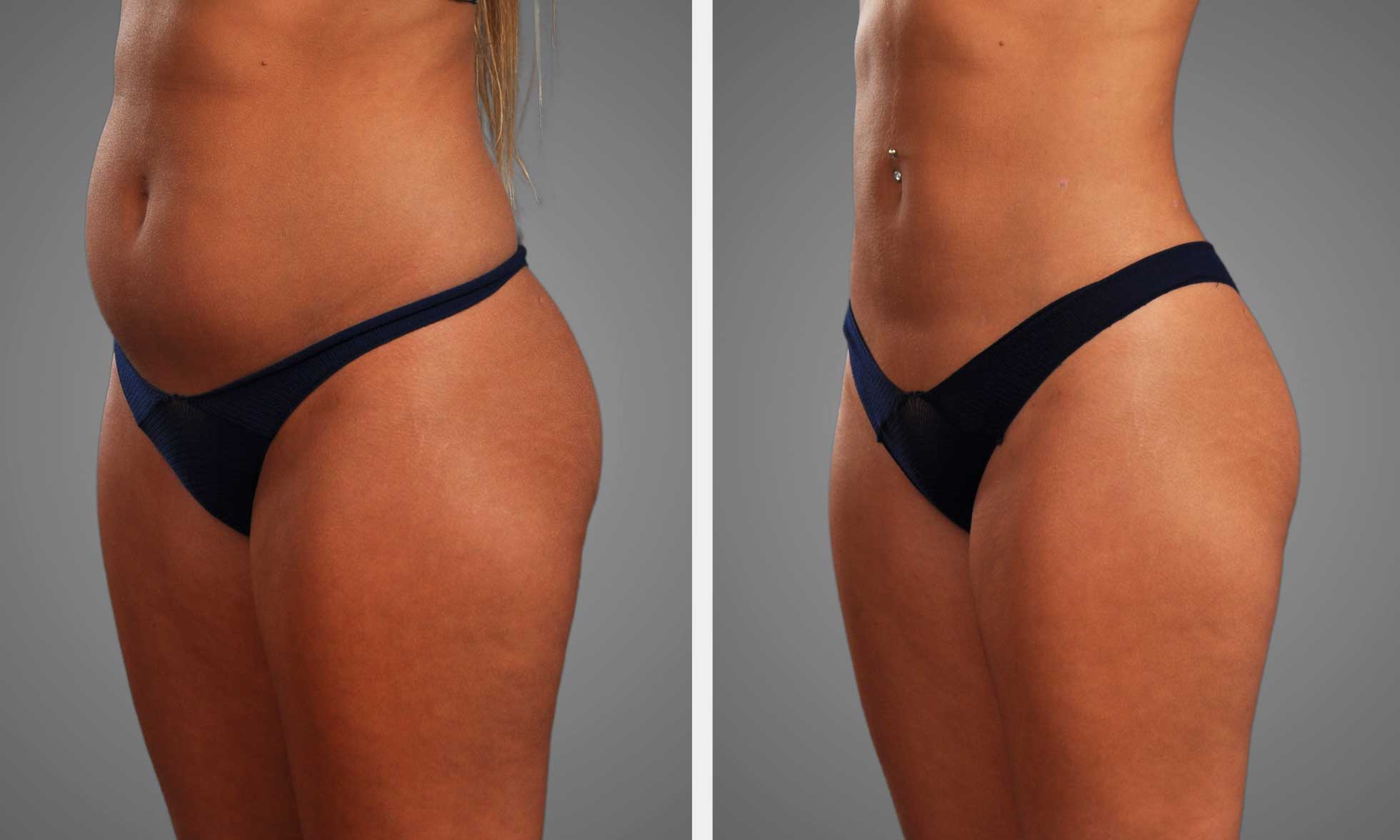
Liposuction, especially water-assisted or tumescent approaches, decreases fat overload and may improve discomfort and functional status. Liposuction could be reimbursable for stage III patients fulfilling local criteria.
Check with your regional health policy and documentation requirements prior to scheduling surgery. Lifestyle measures reinforce any medical approach. Daily low-impact exercise, like walking, swimming, or biking, keeps you mobile and lessens flares.
Track activity using straightforward devices such as pedometers. Skin care avoids infections and preserves barrier function. Anticipate symptom improvement post-treatment, but understand the disease may continue. Strategies have to evolve.
Management options and expected outcomes:
- Complex decongestive therapy provides symptom relief and reduces swelling. It requires ongoing use.
- Compression garments: improved comfort, slow progression; compliance needed.
- Physical therapy and exercise lead to better function and less pain. Long-term habits are required.
- Liposuction (select cases): significant volume reduction, surgical risks and rehab.
Expert Care
Build a team of experts from multiple disciplines. Core members range from vascular specialists and dermatologists to physical therapists and a surgeon with experience in lipedema procedures.
Add a psychologist or psychiatrist for mood, body image, and eating disorder issues. A certified lymphedema therapist does the CDT and garment fitting.
Post-Diagnosis Coordinated care improves outcomes by aligning goals, reducing redundant tests, and timing interventions such as surgery with optimized conservative care beforehand.
Maintain a collaborative care plan with defined responsibilities and consistent check-ins. Provide patients with a short list of key providers: primary care physician, lymphedema therapist, vascular or plastic surgeon, dietitian or psychologist, and physiotherapist.
Long-Term View
Lipedema is chronic and requires life-long management and routine follow-up to track progression and tailor treatments. Organize routine staging reviews and revision activity lists.
Support groups and community resources provide practical tips and emotional support and recommend joining local or online groups. Psychological support aids in depression, anxiety, and dealing with the chronic change.
To remain functional, strategies include ongoing CDT as needed, exercise, and early treatment of skin breaks or infection.
Conclusion
Lipedema requires definitive action and compassionate attention. Clinical exam and patient history are at the heart. Ultrasound checks tissue, and MRI is used for hard cases. Simple measures such as waist-to-hip ratio and limb volume assist in monitoring change. Match diagnostics with patient narratives to complete the profile. Interdisciplinary teams combining physicians, therapists, and nurses yield superior outcomes. Emerging technologies, including advanced imaging and biomarkers, hold promise but require clinical validation. Effective follow-up and self-care regimens reduce swelling and pain and maintain mobility. For anyone concerned about leg or arm swelling, begin with a specialist familiar with lipedema and inquire about imaging, thorough examination, and ongoing care. Book a consult or a second opinion.
Frequently Asked Questions
What is the most reliable method to diagnose lipedema?
Clinical assessment by a specialist (vascular medicine, lymphology, or a dermatologist) combined with medical history and physical exam is the most reliable first step. Imaging like ultrasound or MRI supports the expert clinical diagnosis but does not replace it.
Can imaging tests confirm lipedema?
Imaging (ultrasound, MRI, lymphoscintigraphy) helps rule out other conditions and shows tissue changes. They support diagnosis but do not definitively diagnose by themselves. An expert reads imaging in conjunction with clinical presentation.
How does lipedema differ from lymphedema and obesity?
Lipedema exhibits symmetrical fat deposition in the limbs, pain, and easy bruising. Lymphedema causes fluid swelling and skin alteration. Obesity creates generalized fat without the pain or bruising common in lipedema. A clinician tells these apart by exam and history.
Are there blood tests for lipedema?
There is no blood test for lipedema. Blood work can rule out other causes, such as hormonal or metabolic issues. Diagnosis is based on clinical features and imaging.
When should I see a specialist for suspected lipedema?
Visit a specialist for persistent, painful limb enlargement, disproportionate fat, easy bruising, or poor diet or exercise response. The sooner you get an evaluation, the better your management and symptom control.
How do clinicians stage or grade lipedema?
Clinicians stage lipedema by tissue changes and severity (mild to severe) and measure functional impact. Staging guides treatment decisions such as conservative treatment or surgery options including lipedema-aware liposuction.
What are the next steps after a lipedema diagnosis?
Begin conservative care: compression, exercise, manual lymphatic techniques, and pain management. Explore surgical options and long-term follow-up with a specialist to reduce your symptoms and improve function.