Key Takeaways
- Look out for a soft, persistent lump or growing localized swelling that feels unlike typical postoperative hardness and might signal a seroma.
- Fluid collects, so seromas often become evident by a wave or sloshing sensation under the skin and clear, straw-colored drainage from the incision.
- Pay attention to ongoing tenderness, localized pain, skin changes such as redness or thinning, and delayed healing which can signal a symptomatic seroma requiring evaluation.
- Distinguish seroma from typical healing by persistence in duration, size, and consistency. Seromas tend to be persistent, localized, soft, and related to clear fluid instead of diffuse mild swelling.
- Reduce risk and support recovery by following aftercare. Wear compression garments, limit activity, manage drains properly, and attend follow-up appointments.
- Evaluate with a healthcare provider if symptoms persist or worsen so diagnosis can be confirmed with a physical exam or ultrasound and a personalized treatment plan initiated.
Seroma after liposuction signs are pockets of fluid that develop beneath the skin after an operation.
Typical symptoms are a fluctuant swelling, clear or slightly yellow drainage, warmth without high fever, and a discreet change in contour at the treated site.
Small ones usually resolve with time, while larger seromas may require clinical drainage.
Early recognition assists in directing safe follow-up care and minimizing the risk of infection or extended recovery.
Seroma Signs
Seroma liposuction shows as a collection of serous fluid underneath the skin. Seroma signs normally follow within 5 to 10 days of surgery or drain removal. Patients frequently arrive 7 to 10 days post-op with a soft, fluctuant pocket adjacent to the incision. Early recognition directs treatment decisions like observation, aspiration, or additional testing when infection is suspected.
1. Localized Swelling
Identify unusual or escalating swelling in the vicinity of the liposuction site, which doesn’t decrease with typical recovery interventions. Compare the swollen area to the other side to identify asymmetry or a well-defined lump that indicates a fluid pocket and not just generalized post-operative swelling.

If it feels soft and fluctuates and moves a bit under your fingers, that again indicates seroma. Follow changes over multiple days. Normal post-op swelling will diminish; seroma swelling often does not and may increase. Collections in excess of 75 to 100 ml more frequently cause pain and limited function.
2. Fluid Sensation
Upon palpation of the area, you may experience a wave-like or sloshing sensation beneath the skin, suggesting mobile serous fluid. There may be a shifting or compressible lump versus a firm, fibrous nodule.
Patients may describe a “water balloon” effect: unusual softness and give with pressure. Distinguishing this from solid masses is important because fluid collections often respond to needle aspiration, though aspiration risks infection and for some locations, implant damage or pneumothorax.
3. Clear Drainage
Observe any clear, straw-colored, or lightly yellowish fluid leaking from the incision or wound. This odorless, non-bloody drainage is characteristic of seroma and is not like frank bleeding or purulent drainage.
Note the volume and frequency of leakage. Continuous drainage indicates a persistent cavity. If aspirated fluid is cloudy or purulent, send a sample and consider broad-spectrum antibiotics if infected.
4. Persistent Tenderness
Tenderness or pain at the surgical site that persists is another sign. If pain intensifies with motion or local pressure, think underlying fluid collection. Localized pain confined to one area is more suggestive of a symptomatic seroma than the generalized soreness that occurs after surgery.
Persistent pain with swelling or drainage needs clinical review and possible intervention.
5. Skin Changes
Check for redness, warmth, or shiny stretched skin over indicators of fluid pressure on the skin. Thinning or color change, delayed wound healing, or small areas of skin breakdown can occur secondary to a persistent seroma.
Redness and systemic signs or purulence concern for infection. Don’t delay; management may include drainage, antibiotics, and further evaluation.
Seroma vs. Normal Healing
Some swelling and firmness following liposuction is normal with tissue settling and fluid shift. Early swelling is typically diffuse, symmetric and gets progressively better over days to weeks. A seroma is a localized collection of clear fluid that can develop under the skin or between tissue layers where the lymphatic channels or small blood vessels are inadvertently damaged.
Differentiating seromas from normal healing educates readers on when to reach out to their surgeon and what to anticipate during the initial days and weeks following surgery.
Recognizing the difference
Anticipated post-op puffiness tends to be diffuse and squishy, typically under circumstances where the suctioning was strongest. It shifts in shape throughout the day and with body position, and slowly diminishes over two to six weeks with compression, ambulation, and time.
In comparison, seromas feel like a pocket or lump beneath the skin. They can feel like a soft, spongy area that gives when touched, and occasionally a fluctuance can be palpated. Seromas can actually expand instead of contract in the days following surgery and cause local tension or slight pain without the widespread bruising that accompanies normal trauma.
Key differences in symptoms:
- Duration: Normal swelling peaks within 48 to 72 hours and slowly improves over weeks. Seromas can continue or arise after week 1 and stay the same or grow.
- Size: Typical postoperative swelling is diffuse. Seromas form discrete pockets that can be as small as beans or gums to a few centimeters across.
- Consistency: Normal swelling feels firm or puffy. Seromas are soft and fluctuant, sometimes shifting under pressure.
- Location: Normal edema follows the treated area broadly. Seromas are isolated in one area or within incision lines.
- Fluid character: Seromas usually contain clear, straw-colored fluid. Hematomas are dark and clotted, and normal swelling has no discrete fluid pocket.
- Symptoms: Seromas can cause localized tightness, discomfort, or mild skin blanching. Infection would cause additional redness, worsening pain, heat, or bad discharge.
Seromas often exhibit clear fluid and local signs as opposed to diffuse, mild post-op changes. For instance, an abdomen liposuction patient who observes a focused, soft bump just beneath the right incision that feels like a water balloon probably has a seroma, while generalized belly swelling that dissipates with compression is likely normal healing.
Some small seromas simply resolve on their own with compression and movement. Larger or persistent pockets might require needle aspiration in the clinic or drain placement to prevent ongoing accumulation and infection risk.
Track changes, timing and symptoms, and any fever, increasing pain, or foul discharge to a provider as soon as possible.
Underlying Causes
Seroma after liposuction develops when lymphatic vessels and tissue planes are damaged during the procedure, enabling serous fluid to accumulate in the void. Liposuction separates fat from surrounding tissue with cannulas, and this physical disruption often transects small lymphatic channels. When those channels are unable to drain normally, fluid builds up under the skin.
This is a wound-healing problem tied to the first phase of repair. A prolonged early phase with slowed angiogenesis and epithelialization makes the area heal more slowly and lets fluid persist rather than resolve quickly.
Excessive tissue manipulation increases the risk. Working over large surface areas or making multiple passes with the cannula both increase soft tissue trauma and generate additional dead space. Removing such vast quantities of fat in a single sitting boosts this impact.
For instance, working multiple areas, such as the abdomen, flanks, and thighs, in the same procedure generates large surfaces for fluid to collect. There is the same worry when surgeons employ aggressive suctions or wide dissections. More raw surface leads to more exudate and seroma risk.
Poor closure of tissue spaces is another cause. When the planes of tissue are not re-approximated either because quilting sutures are not placed or because Scarpa’s fascia is not preserved, pockets exist where fluid can accumulate.
Quilting sutures and preservation of Scarpa’s layer have decreased seroma rates in many centers as preventative steps. Closed-suction drainage additionally assists by removing fluid as it forms, so not placing drains when indicated or placing too few leaves patients vulnerable.
Improper use or early drain removal is the most common cause of seromas. Drainage tubes are intended to decrease dead-space collections until adherence of tissue and lymphatic repair. Taking drains out before production slows to an appropriate rate can lead to a seroma forming.
Fluid accumulation following premature removal is a common precipitating factor. High-volume seromas, above roughly 75 to 100 ml, are more prone to pain, infection, and compromised function of the treated site, so timely management counts.
Other contributors are damage to underlying structures during dissection. Nerves, vessels, and even tendons can get damaged, exacerbating local irritation and fluid production.
Periprosthetic seromas have implant damage in them, and chest wall seromas have a pneumothorax, each demonstrating how diverse the local causes can be. Tissue adhesives won’t reliably prevent seromas but may reduce their volume.
While breast surgery has been studied the most, seroma formation is not completely understood and can occur after any operation that involves extensive soft-tissue dissection.
Patient Risk Factors
There are patient factors that determine how prone you are to seroma formation after liposuction. These influence tissue reaction, lymphatic circulation, and the rate of healing. Knowing them allows clinicians to predict risk and customize consent, technique, and follow-up.
Procedure Extent
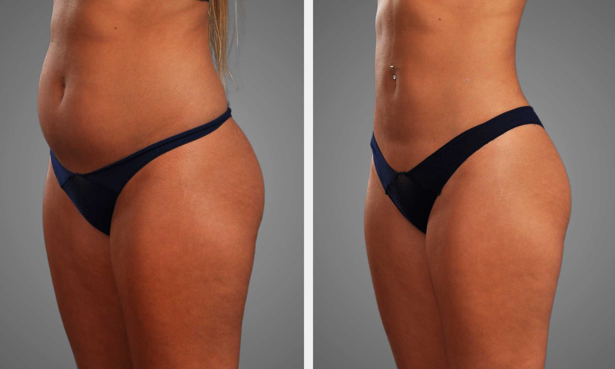
Bigger or more extensive liposuction raises seroma risk since more fat and tissue are removed, creating larger potential spaces for fluid to accumulate. Treating multiple sites in the same session adds up those voids and your body might not reattach tissues quickly enough to avoid fluid pools.
High-volume fat removal, usually considered to be multiple liters, increases the risk of seroma compared to small, targeted liposuction. Pairing liposuction with another major surgery, like abdominoplasty, multiplies risk. The additional dissection and undermining further destroy lymph channels and establish larger dead spaces.
By tracking the size and number of treated areas, teams can anticipate which patients are at risk for requiring drains, extended compression, or closer follow-up. Surgeons have some control over risk. They can stage procedures so that a smaller area is treated during a single session or use techniques that reduce tissue trauma.
Recording area size, aspirate volume, and operative time comes in handy for postoperative planning.
Medical History
Seroma, hematoma, and other wound complications in the past obviously foreshadow higher recurrence risk. Previous tissue behavior to fluid predicts a lot of future risk. Certain chronic illnesses like diabetes will impact your healing and therefore increase the risk of seroma because elevated blood sugar slows tissue repair and alters immune function.
Autoimmune disorders can likewise change inflammation and impede normal fluid clearance. Previous surgeries in the same area, such as hernia repair, C-section, or prior liposuction, can scar or disrupt lymph channels. That disruption makes drainage less efficient and encourages fluid accumulation.
Medications matter: anticoagulants increase bleeding and hematoma risk, which can progress to seroma. Systemic steroids depress immunity and slow healing. Certain pills might bump it up a bit; stopping them around two weeks pre-surgery is generally recommended.
Age and general health matter: older or frail patients usually heal slower and can be more susceptible.
Aftercare Compliance
Nice post-op care diminishes the seroma rates. Properly used compression garments support tissues and restrict room for fluid accumulation, while improperly wearing or not wearing them at all increases your risk. Any activity restriction and heavy lifting avoidance prevent shearing forces that may reopen space or displace tissue layers.
Wound care and follow-up are important. No-shows postpone early fluid collection detection. Proper drain management when drains are employed reduces seroma formation. Drains should be emptied and removed as per protocol to prevent infection or persistent tracts.
Checklist:
- Wear a compression garment full-time as directed, usually for weeks.
- Do not do heavy lifting or vigorous exercise until cleared by a clinician.
- Keep incision sites clean and dry. Notice increased swelling or drainage.
- Write and bring drain output logs to follow-up visits.
- Quit smoking a minimum of three weeks prior to surgery, with some meds to stop two weeks prior when indicated.
The Diagnostic Journey
Seroma post-liposuction is a clinical diagnosis starting with directed history and physical examination, followed by directed imaging and differential diagnosis to guide care. Early recognition is important because seromas can alter the speed and course of recovery and impact future treatments.
Begin with symptom and timing reported by the patient. Inquire when swelling or strange sensations commenced. Seromas usually develop approximately 7 to 10 days post surgery but may present weeks or even months later. They usually present with patients who describe a soft bulge, a feeling of fluid shifting, mild tightness, or a new area of fullness that is different from expected post-op swelling.
Observe if there is any variation with motion or position and if pain, erythema, or heat is present. Conduct a targeted physical examination of the surgical site. Palpate for soft, fluctuant swelling and check both sides. A mere tap to one side reveals the tell-tale wave motion under the skin that betrays a fluid pocket.
Measure the size and location, and record the findings. Examine skin color, warmth, and skin breakdown. Record any incisional drainage or systemic signs that indicate something beyond a sterile seroma.
Employ ultrasound as the first line of imaging to confirm fluid and to estimate volume. Ultrasound is broadly available, noninvasive, and sensitive to fluid collections. It demonstrates anechoic or hypoechoic pockets compatible with seroma and permits the provider to quantify depth and volume in milliliters.
Imaging helps direct care decisions between conservative care and aspiration and can monitor resolution through follow-up visits. Distinguish seroma from hematoma, abscess, and lymphedema. Hematomas present earlier, with firmer, sometimes tender masses and evidence of blood on imaging.
Abscesses demonstrate thicker walls, internal echoes on ultrasound, and systemic signs such as fever and leukocytosis. Lymphedema is often diffuse and chronic, not well localized to a single pocket. Proper diagnosis keeps you from the wrong treatment and potential complications.
Schedule diagnostic and next steps based on results. Small, asymptomatic seromas may be observed with compression and follow-up ultrasound. Larger or symptomatic seromas might require needle aspiration with appropriate sterile technique.
Record the volume removed and submit fluid to the lab for analysis if infection is suspected. Seromas that are recurrent or loculated may need imaging-guided drainage or surgical revision. Always advise patients that duration varies; some resolve in weeks, while others take months.
If a seroma is suspected, consult a qualified healthcare provider for evaluation and clear guidance on management and monitoring.
Treatment Options
Treatment for post-liposuction seroma ranges from conservative management to operative intervention to drain the fluid, relieve the pain, and avoid infection or recurrence. Selection of strategy varies based on seroma size, location, time frame, symptoms, and the patient’s requirements.
Small collections can resolve as the body reabsorbs fluid over several weeks. Larger, symptomatic, or recurrent seromas require intervention.
Conservative Management
Surgical drains that are removed when the output falls below a certain level help pull the fluid out of the body. Follow garment fit and cleaning guidelines. Badly fitting garments can exacerbate swelling.
Warm compresses for short sessions can help natural drainage and reduce swelling. Apply with a warm cloth to prevent scalding. Rest and elevation of the affected area decrease venous pressure and enhance fluid resorption.
Restrict heavy lifting and strenuous activity until you have been cleared. Movement that stresses tissue can exacerbate seromas.
Manual lymphatic drainage (MLD) by a trained therapist can accelerate fluid movement and reduce the risk of recurrence. MLD is a soft, focused massage that encourages circulation in the region.
Techniques to manage seromas:
- Wear surgeon-prescribed compression garment consistently.
- Use warm compresses two to three times daily for ten to fifteen minutes.
- Elevate and rest the area and avoid lifting heavy objects.
- Book MLD sessions with a certified therapist when advised.
- Monitor skin for warmth, redness, or increasing pain.
Aspiration
Needle aspiration is a minimally invasive method of draining seroma fluid when it becomes painful or functionally limiting. Most frequently, it is done in-office under sterile conditions with a small needle and syringe.
This typically allows for rapid symptom resolution and can facilitate walking if the collection was impeding that. Certain seromas refill after aspiration, requiring multiple taps.
If refilling is frequent, explore small surgical options to reduce infection risk from repeated needle sticks. Once aspirated, observe for infection and schedule follow-up visits to monitor recurrence.
Surgical Intervention
Surgery is for large, stubborn, or complex seromas that don’t respond to other treatment. The surgery might open the seroma cavity, excise fibrous tissue, and insert a drain for ongoing fluid drainage.
Drains are usually left in for a few days to a week and minimize new fluid accumulation. Surgical objectives are eliminating persistent pockets and returning normal tissue contour.
Postoperative care is critical. Wound checks, proper drain care, and adherence to activity limits reduce recurrence. With infection or complicated scarring, tissue removal might be required to avoid chronic issues.
Conclusion
Seroma after liposuction manifests itself by soft swelling, fluid shift, and a mild tug on the skin. Brief checks for warmth, increasing size, or discoloration indicate an issue. Clear fluid on the dressing or a sudden bulge signal the necessity for a medical check. Early diagnosis is done with an exam and simple imaging. Small seromas may drain by themselves or with needle aspiration. Larger or persistent collections require directed drainage and short-term compression. Risk increases with large amounts of tissue manipulation, some medical problems, and inadequate post-care.
Simple steps help lower risk: follow wound care, wear compression, rest from hard strain, and keep follow-up visits. Consult your surgeon at the initial indication of alteration.
Frequently Asked Questions
What is a seroma after liposuction?
Liposuction seroma is a pocket of clear fluid that builds up under the skin after liposuction. It develops where tissue was dissected or excised and can be doughy, edematous, or flutuant.
How quickly do seroma signs appear after surgery?
Seroma symptoms generally present themselves within days to a few weeks post-surgery. Some develop later, but the majority present during early recovery when swelling starts to fluctuate.
How can I tell a seroma from normal postoperative swelling?
A seroma is a localized, ‘soft’ pocket of fluid that can shift with pressure. Normal swelling is more diffuse and firm. If you detect a distinct bulge or fluid wave, call your surgeon.
What are the main risk factors for developing a seroma?
Risk factors comprise large-volume liposuction, multiple areas treated, extensive tissue dissection, obesity, and postoperative ambulation or suboptimal compression garment wearing.
How is a seroma diagnosed?
Diagnosis is by clinical exam supplemented by ultrasound when necessary. Ultrasound identifies the size of the fluid collection and guides drainage.
When does a seroma need treatment?
Manage seroma if it results in pain, significant bulging in the area, infection risk, or impedes wound healing. Small, asymptomatic seromas can spontaneously resolve.
What are common treatments for a seroma after liposuction?
Treatments include needle aspiration, repeated drainage, pressure dressings, compression garments, and rarely surgical drainage or placement of drains if persistent or infected.