


Key Takeaways
-
Liposuction can cause post-surgical hyperpigmentation through inflammation, hemosiderin deposition, and tissue trauma. Anticipate temporary or occasionally persistent skin discoloration post-procedure.
-
Evaluate individual risk by considering skin type, genetics, and medical background. Individuals with darker complexions and a history of pigmentary disorders have an elevated risk of developing postinflammatory hyperpigmentation.
-
Do what you can before and after surgery. Avoid tanning, use the topical preparations prescribed when indicated, and adhere to wound care and sunscreen regimens to minimize pigment issues.
-
Keep a close eye on healing and maintain a mini checklist of post-op care: daily broad-spectrum sunscreen, gentle moisturizers, and avoiding harsh exfoliation or irritants.
-
For stubborn or intense hyperpigmentation, pair topicals with in-office procedures such as lasers or chemical peels under dermatological supervision. Be sure to let your skin heal before aggressive treatments.
-
Surgeons should maximize technique, incision location, and patient selection. They should convey reasonable timelines and the potential for staged touch-ups to limit hyperpigmentation.
Hyperpigmentation after liposuction treatment is a frequent skin reaction in which treated zones become darker from excess melanin. It tends to show up a few weeks post surgery, impacting the look of healing, tone and scar visibility.
Risk increases with deeper procedures, inflammation or sun exposure. Prompt aftercare, mild skincare and sun avoidance decrease the risk of permanent discoloration.
The sections below discuss causes, prevention measures, and actionable treatments for post-liposuction pigmentation.
Understanding The Cause
Liposuction can trigger a cascade in the skin that results in brownness. Surgical trauma, blood staining, and inflammation damage the dermis and subcutaneous layers. That injury starts local healing signals and an increase in melanin. Pigment can migrate as tissue remaps during recovery, leaving patches, splotches, or diffuse discoloration that can persist for months or years.
1. Inflammation Response
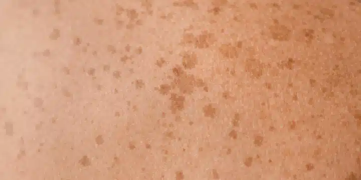
Tissue injury during liposuction induces an inflammatory cascade. Immune cells flood in, cytokines soar, and local melanocytes are provoked to produce additional melanin pigment. This process results in postinflammatory hyperpigmentation (PIH) which appears as brown or gray-brown spots in the areas of skin that were stressed.
Ongoing redness or swelling continues to stoke the inflammatory signals, which can turn a temporary dark spot into a long-term patch. Slow or complicated wound healing caused by infection or poor circulation keeps melanocytes going longer and raises the risk of persistent pigment alteration.
2. Blood Pigments
Bruising leaves blood breakdown products in the upper layers of the skin. Hemoglobin from red blood cells degrades into hemosiderin and other pigments that discolor tissue brownish or rusty. They can resemble irregular splotches or individual compacted dark spots, depending on the severity and spread of bruising.
Because of slow healing and persistent edema, blood pigments are allowed to linger and embed, making the discoloration more persistent. Where blood pools under thin skin, the effect is more dramatic and may fade only gradually over months.
3. Tissue Trauma
Surgical trauma ruptures dermal cells and the dermal connective matrix, and can distort the normal architecture that holds pigment uniform. Deeper wounds, poor skin retraction, or necrosis can leave behind permanent pigment changes or scarring that changes color and texture.
Aggressive or repeat suctioning leads to a higher risk of abnormal scarring patterns and new pigmented lesions. Existing conditions like melasma or a history of PIH make this more likely, and postoperative infections can exacerbate discoloration even more.
4. UV Radiation
Sun exposure following surgery accelerates melanin activity and can transform a temporary pigment shift into a permanent issue. UV light intensifies inflammation induced melanin production and hyperpigments both PIH and blood-stain regions.
Apply broad-spectrum sunscreen with a high SPF and cover treated areas. Don’t tan intentionally. Protection is particularly crucial in these first months when skin is remodeling and is most sensitive.
5. Surgical Technique
Technique matters: Tumescent methods, cannula size, and incision placement affect tissue trauma and thus pigment risk. Longer procedures or bad technique increase the risk of necrosis and pigment migration.
Thoughtful incision siting, staged treatments, and clear wound-care instructions minimize chronic discoloration. Laser, peel, and topical therapies can assist afterward, but response depends on skin type and pigment.
Assess Your Risk
Assessing the likelihood of hyperpigmentation after liposuction starts with a clear look at three areas: skin type, genetic factors, and medical history. Each affects how skin responds to trauma, how much melanin is produced during healing, and how long discoloration may last. Use the checklist and guidance below to judge individual risk and set realistic expectations.
Skin Type
More of a risk with darker skin tones, such as Fitzpatrick type V, is postinflammatory hyperpigmentation. Melanin-rich skin responds to surgical trauma with aggressive pigment production. A small bruise or surface inflammation can leave brown or gray patches that come through more clearly on brown and dark skin.
Watch for uneven tone, new brown spots, or persistent darkening in the weeks following surgery. Hyperpigmentation tends to surface within weeks to a few months, and mild cases respond well to care and subside in three to twelve months.
Variations from fair to very dark skin influence visibility. Lighter skin may show pink or red healing but less lasting brown pigment. Mid-tones can develop patchy brown areas, and darker tones often show denser, longer-lasting discoloration.
Inform your patients with richer tones that they need to anticipate closer follow-up and interventions such as rigorous sun protection and early topical management.
Genetics
If you have a family history of pigmentation issues, your risk goes up. Genetic predispositions impact inherent melanin levels, the speed at which pigment cells activate post trauma, and scarring trends.
Here is a basic chart of genetics and average impact on pigment shift.
|
Genetic factor |
Typical effect on pigment |
|---|---|
|
Family history of hyperpigmentation |
Higher chance of post-procedure darkening |
|
Tendency to keloid or hypertrophic scars |
Uneven tone and persistent patches |
|
Ethnic background with high melanin |
Faster, deeper pigment response |
Inherited issues like melasma or postinflammatory predilection can result in lingering, patchy tone following liposuction. Surgeons should inquire explicitly about relatives with pigment problems to improve risk predictions.
Medical History
Screen for previous pigment problems, eczema, scarring and any dermatologic diagnoses prior to liposuction. Chronic inflammatory skin disease, prior laser or chemical treatments and a history of extended post-treatment discoloration all alter the risk.
Detail any previous cosmetic procedures and topical or oral skin treatments. Past bruising and scar tendencies are indicative of future healing.
Keep in mind that altered pigmentation, bruising, and scar types typically resolve within weeks to months, but certain pigment alterations may persist for months or years. Advise patients to protect healing skin from UV exposure, utilize broad-spectrum sunscreen, and that preemptive care and setting expectations are everything.
Proactive Prevention
Hyperpigmentation prevention after liposuction is a combination of pre- and post-surgery steps that reduce inflammation, protect skin barrier function, and minimize pigment activation. It’s about reducing risk factors, promoting wound repair, and early problem detection.
Pre-Surgery
Prime skin with topicals that even tone and inhibit melanin production like azelaic acid and hydroquinone when indicated or gentle retinoids with clinicians input. Proactive prevention is essential. Short supervised superficial chemical peels, such as glycolic or lactic at low strength, help trim patchy tone if done weeks pre-op.
Discontinue retinoids about seven days pre-op or as directed to avoid fragility. Avoid tanning, sunbeds, or intentional UV exposure for a few weeks pre-op because UV activates melanocytes and increases the risk of PIH. No aggressive scrubs or exfoliation right near surgery; they cause micro-injury and inflammation.
Document baseline skin findings with dated photos and a brief history of previous pigment issues, hormonal factors, or photosensitizing medications. This assists clinicians in benchmarking outcomes and making early interventions. Talk about a customized plan if you have Fitzpatrick skin types IV–VI, a history of melasma, or post-inflammatory hyperpigmentation tendency.
This might involve pre-op topical suppression, altered surgical technique, or staged procedures.
Post-Surgery
If wounds are present, clean wound sites gently with mild, non-foaming cleansers, keep skin dry when instructed, and use sterile dressings as directed. Use broad-spectrum sunscreen (SPF 30 or more) on any exposed areas once wounds have closed and for every day thereafter. Sunscreen is essential because it prevents UV-driven exacerbation of any new discoloration.
Moisturize with gentle fragrance-free creams to maintain supple skin and help repair the barrier. Initiate light emollients as soon as dressings are removed and increase as advised by your clinician. Wear compression garments as advised, typically 24/7 for a few weeks and as long as six weeks for optimal skin retraction to mitigate fluid accumulation, support shape, and minimize the risk of issues that cause pigmentation.
No strenuous activity for approximately two weeks. Limited movement decreases bleeding, swelling, and the risk of seroma. Watch incision sites and treated areas for black spots, delayed tanning, or stubborn marks. Seromas are detected early and, if needed, drained. They can prevent lingering inflammation that results in residual hyperpigmentation.
Refrain from aggressive topicals, strong acids, or retinoids until the skin has completely healed and your clinician clears them.
-
Preventative measures by risk and skin health:
-
High-risk pigment history: pre-op topical suppression, close follow-up, staged treatment.
-
Recent tanning: delay surgery until fade and ensure strict sun avoidance.
-
Sensitive or dry skin: start gentle moisturizers weeks before surgery.
-
Post-op fluid risk: strict compression garment use for the recommended duration.
-
Activity level: rest and avoid heavy exertion for two weeks.
-
Long-term: periodic check-ins every few months for early issue detection.
-
Corrective Treatments
Post liposuction hyperpigmentation can be addressed with everything from daily topical care to in-clinic procedures. Selection is based on pigment depth, skin type, and duration of discoloration. Permanent alterations occur in around 6 to 10 percent of patients; therefore, a structured, gradual approach aids in establishing reasonable expectations.
Multiple treatments are usually required to achieve an even tone, and many clinicians recommend waiting 6 to 12 months post surgery before initiating aggressive correction.
Topical Agents
-
Hydroquinone (often prescription-strength for stubborn patches)
-
Azelaic acid
-
Kojic acid
-
Vitamin C serums (topical antioxidants)
-
Retinoids (nightly adapalene, tretinoin or similar)
-
Mild corticosteroid creams (short term, under guidance)
-
Brightening combinations (e.g., hydroquinone plus retinoid and sunscreen)
Prescription topicals are the go-to once OTCs fall short. Stubborn brownish round patches typically require hydroquinone or higher-strength retinoids under a dermatologist’s supervision. Topicals reduce melanin synthesis and seep out pigment over time.
Results might manifest within weeks for certain agents, while maximal benefit may require months. Consistent use is what counts. A standard schedule combines a retinoid at night with an antioxidant serum and broad spectrum SPF 30 plus during the day.
Watch for irritation; discontinue or lessen strength if redness or peeling develops. Patch testing and slow titration mitigate the risk of sensitization.
Professional Procedures
Laser and light therapies, chemical peels, and deeper resurfacing are options for more severe or deep set pigment. Energy-based treatments such as Nd:YAG, excimer, and narrowband UVB can work well for deeper pigment. They usually require multiple sessions and may show improvement after a few treatments.
The full course typically extends to 10 treatments depending on response. Chemical peeling ranges from superficial glycolic or lactic peels to medium-depth trichloroacetic acid peels. Deeper peels can address stubborn discoloration but have higher downtime and risk.
Camouflage tattooing and scar revision may be options when pigment tracks scarred tissue or when color match is possible. Schedule is significant; most recommend waiting six to twelve months before revision surgery or aggressive laser to allow natural fading and scar maturation.
If dark spots persist beyond six months to a year, then staged intervention from a dermatologist or aesthetic surgeon is reasonable.
Home Care
Daily sunscreen and gentle moisturizers guard healing skin and keep more pigment at bay. Choose gentle cleansers and steer clear of scrubs or aggressive exfoliation, which can exacerbate discoloration. Cool masks or formulas with niacinamide and panthenol can calm inflamed skin and assist with visual evening.
Photograph your progress every 4 to 8 weeks to gauge treatment impact and fine-tune regimens. For corrective treatments, if you stall, step up to the pros in consultation with a doctor from there.
The Healing Timeline
Liposuction healing is quite predictable. Pigment changes transition from early redness to darker brown marks and then fading. In the initial days to two weeks, the treated area is frequently red and bruised as blood settles and tiny vessels heal. Early redness is inflammation and swelling, not pigment change. Most patients see visible improvement in swelling within the first few weeks as fluid shifts and the body clears blood products.
By four to six weeks you will notice the initial defined contour alterations and a tone shift. Bruises that made you blue or purple typically turn brown as hemoglobin breaks down. This brown staining is a typical early form of hyperpigmentation and tends to become more apparent as swelling subsides. Most patients say the color noticeably softens by eight to twelve weeks, though change is not instant and can take that full period before you see consistent fading.
Between three and six months, the bulk of the healing and skin settling takes place. Most swelling improves by three months, and most color changes have faded in that timeframe. Visible contour changes keep refining, and areas that appeared patchy or uneven in tone typically begin to fade into surrounding skin. Even so, mild residual swelling can persist much longer, often lasting 8 months to a year, which can prevent pigment and contour from settling into its final appearance during that period.
Certain pigment changes outlast the usual timeline. Most discolouration will fade within 2 to 12 weeks, but stains and deeper brown spots can persist and require targeted treatment with topical lighteners, chemical peels, laser treatments or microneedling to improve. It can take many months and sometimes as long as 18 months for skin pigmentation to fully resolve. Schedule follow-ups and multiple treatments if pigment doesn’t fade as anticipated.
Healing areas often appear discolored and mottled in hue before settling. Anticipate uneven patches and shifting tones as the swelling and bruising dissipate and new collagen begins to develop. Factors that impact your healing timeline include your skin type, how deep the fat removal was, how prone you are to bruising, and sun exposure. Shield treated skin from the sun and maintain your provider’s care regimen to accelerate healing and minimize permanent pigment alteration.
|
Stage |
Typical timing |
What to expect |
|---|---|---|
|
Early redness/bruising |
Days to 2 weeks |
Red, blue, purple hues from swelling and blood |
|
Brown staining |
2–12 weeks |
Hemoglobin breakdown leaves brown patches |
|
Contour change visible |
4–6 weeks |
Shape refines as swelling drops |
|
Major settling |
3–6 months |
Most swelling and tone improvement |
|
Long-term resolution |
Up to 18 months |
Residual pigment may fade slowly; may need treatment |
A Surgeon’s Perspective
One surgeon’s take on the hyperpigmentation after liposuction prevention, realistic expectations and early intervention. Preoperative evaluation, surgical decisions, intraoperative methods, and postoperative care all contribute to risk and result. Established truths and standard procedures assist in reducing the risk of permanent discoloration and steer judgment when problems arise.
Patient selection and preoperative assessment matter most. Identify skin type and prior history of hyperpigmentation. Patients with higher Fitzpatrick types are at greater risk of post-inflammatory hyperpigmentation. Review medications, photosensitivity history, and recent tanning or procedures.
Maximize skin health for weeks before surgery by stopping irritant topical agents, treating active inflammation, and ensuring good nutrition and glycemic control. Inform patients that bruising, discoloration, and scar variation can occur and often subside over weeks to months. Sun exposure drives worse outcomes, so advise strict sun block for a minimum of six months after surgery.
There are certain surgical technical choices that can be made that minimize the pigmentary changes. Employ delicate cannulation, smaller cannulae if possible, and refrain from gratuitous subdermal trauma that stimulates inflammation. Use suction strength and make aggressive passes in thin dermis areas.
For more fibrotic tissue, schedule staged sessions instead of hard single-stage removal. Use careful hemostasis to minimize ecchymosis and close with dressings that reduce shear. Keep examples in mind: on inner thighs use smaller cannula and slow passes; around the flanks moderate power with careful tunneling lessens bruising.
Revisions and second surgeries need special care in management. Wait the suggested 6 to 12 months for the tissues and pigment to settle before contemplating revision surgery. Reevaluate skin tone, scar maturation and any residual hyperpigmented patches.
In revisions, think less aggressive aspiration, potential additional skin treatments at the same time and gentle undermining to prevent repeat inflammation. Offer laser options such as the 1,064-nm picosecond-domain Nd:YAG to fade dark spots and even tone when appropriate and space such treatments to avoid overlapping inflammation.
Good communication and organized follow-up is the key. Manage expectations around how long color changes could take and what treatments might be necessary. Take a plain clinic table and very quickly capture skin discoloration information longitudinally – location, size, color, onset, and response to therapy.
Check-ins every few months catch problems early and help adjust therapy. Early topical measures, sun avoidance, or procedural treatments change the trajectory. If pigment changes continue following conservative management, talk about revision surgery as an option beyond the 6 to 12 month mark.
Conclusion
Hyperpigmentation after liposuction doesn’t have to linger. Hyperpigmentation after liposuction treatment Early care and consistent sun protection reduce the risk of dark spots. Watch your skin for gradual color changes and have a clinician examine any new spots. Topical creams, laser touch-ups and microneedling help fade spots in many cases. Most of the change will manifest in weeks to months, not days. A surgeon’s obvious strategy and aftercare accelerate healing and decrease danger. For those considering liposuction, come prepared with your medications, any history of skin reactions, and a candid discussion about your expectations and skin tone. Need assistance selecting a safe next step or clinic question list? Contact me and I’ll get you ready.
Frequently Asked Questions
What causes hyperpigmentation after liposuction treatment?
Hyperpigmentation tends to be a response to inflammation, damage to skin cells, bruising, or melanocytes responding to injury. Sun exposure post-surgery darkens treated areas.
Who is most at risk for post-liposuction hyperpigmentation?
Those with darker pigmentation, a history of post-inflammatory hyperpigmentation, or previous skin conditions are at risk. Cigarettes and some medications can add to risk.
How soon will hyperpigmentation appear and how long does it last?
It usually plateaus within weeks post surgery. Mild ones may dissipate in three to twelve months. Darker or untreated pigment may persist longer or become lifelong without treatment.
What preventive steps reduce the chance of hyperpigmentation?
Avoid the sun, apply broad-spectrum sunscreen (SPF 30+), adhere to your surgeon’s wound-care instructions, and stay away from aggressive skin treatments during recovery. Early anti-inflammatory attention is helpful.
Which treatments improve hyperpigmentation after liposuction?
Topical lighteners (hydroquinone alternatives), peels, laser, and microneedling can assist. Treatment selection varies with skin type and depth of pigment. Seek a skilled dermatologist or surgeon.
When should I see a specialist about persistent pigmentation?
Consult a board-certified dermatologist or your surgeon if pigmentation persists past 6 to 12 months, worsens, or impedes your daily life. Early evaluation allows for more effective, safer treatments.
Can post-liposuction hyperpigmentation be prevented completely?
Full prevention isn’t assured, particularly for darker skin. Risk can be significantly reduced with appropriate pre- and post-op care, sun protection, and immediate treatment of inflammation.
